#
Key functionality
The Relu® Hosted engine offers a wide set of (AI-based) automations based on your input data (medical scans).
#
Input modalities
The following input modalities are supported.
Cone-Beam CT-scans; typically DICOM files. 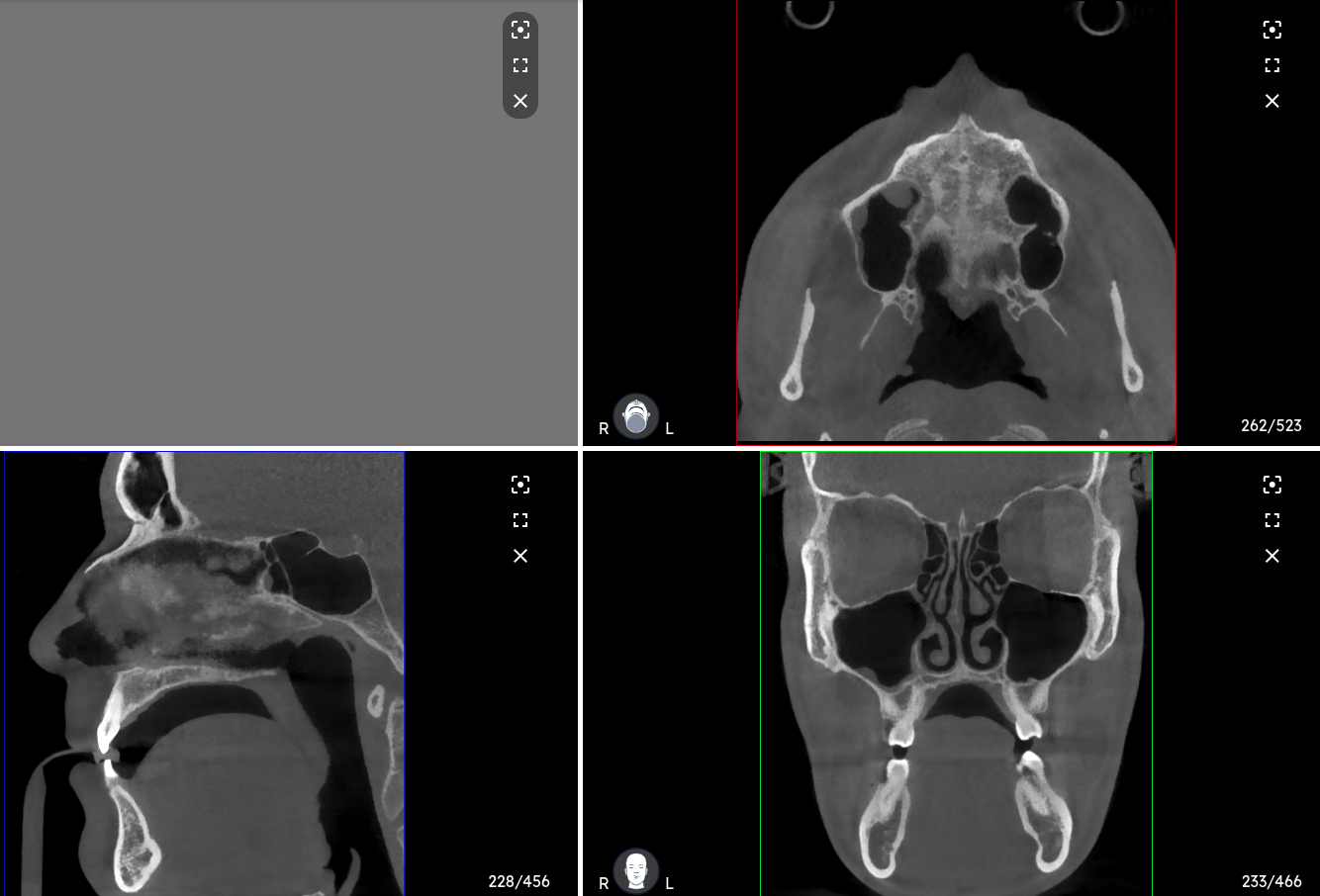
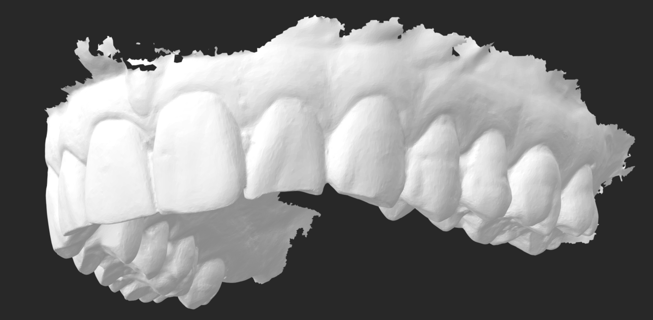
Intra-oral scans or digital impressions for the upper and lower jaw; typically mesh files in .stl, .ply or .obj format.
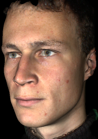
Facial scans; typically mesh files in .stl, .ply or .obj format.
#
Automated results
Based on the input data mentioned above, the Relu® Hosted Engine is able to automatically compute the following outcomes.
Automatic segmentation of the following structures on the Cone-Beam CT-scan:
- full dentition (all permanent and deciduous teeth)
- mandibular bone
- mandibular canals (nerve canals)
- maxillary bone (skull or maxillary-complex)
- maxillary sinuses
- pharynx (airway)
- soft tissue
- root canals
Automatic segmentation of the following structures on the Intra-oral scan:
- dentition (all permanent teeth)
- gingiva (gum), optionally with closed base or emergent profile (
see section below ) - teeth landmarks: axis, center, mesial and distal points (
see section below )
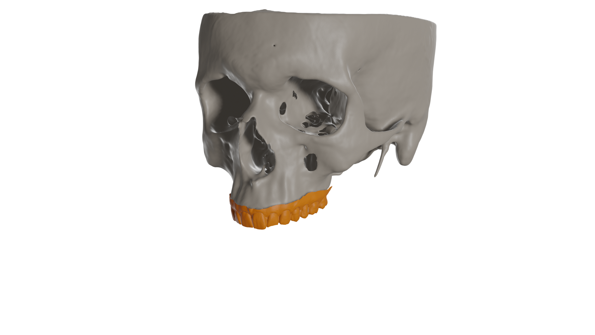
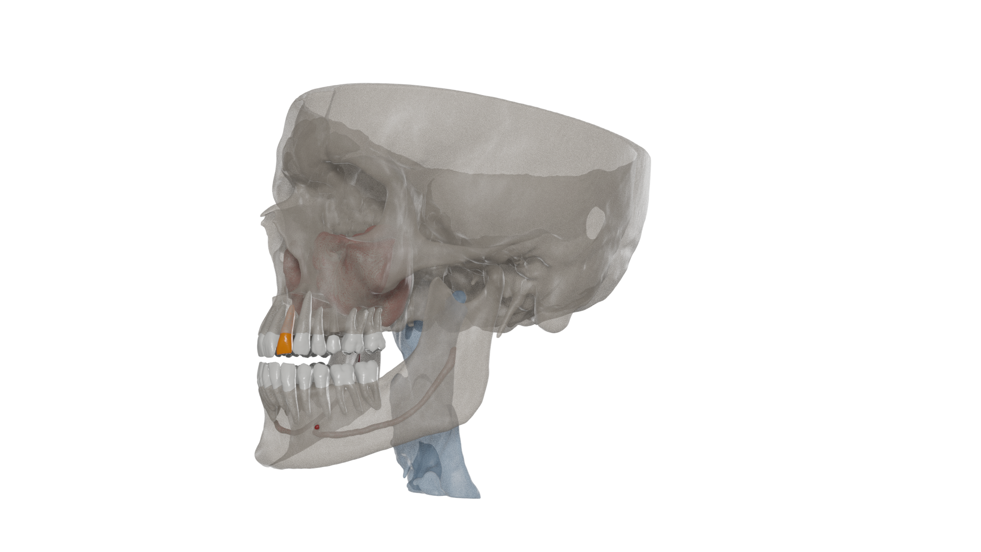
Automatic registration (matching / alignment) of the Intra-oral scan to the Cone-Beam CT-scan.
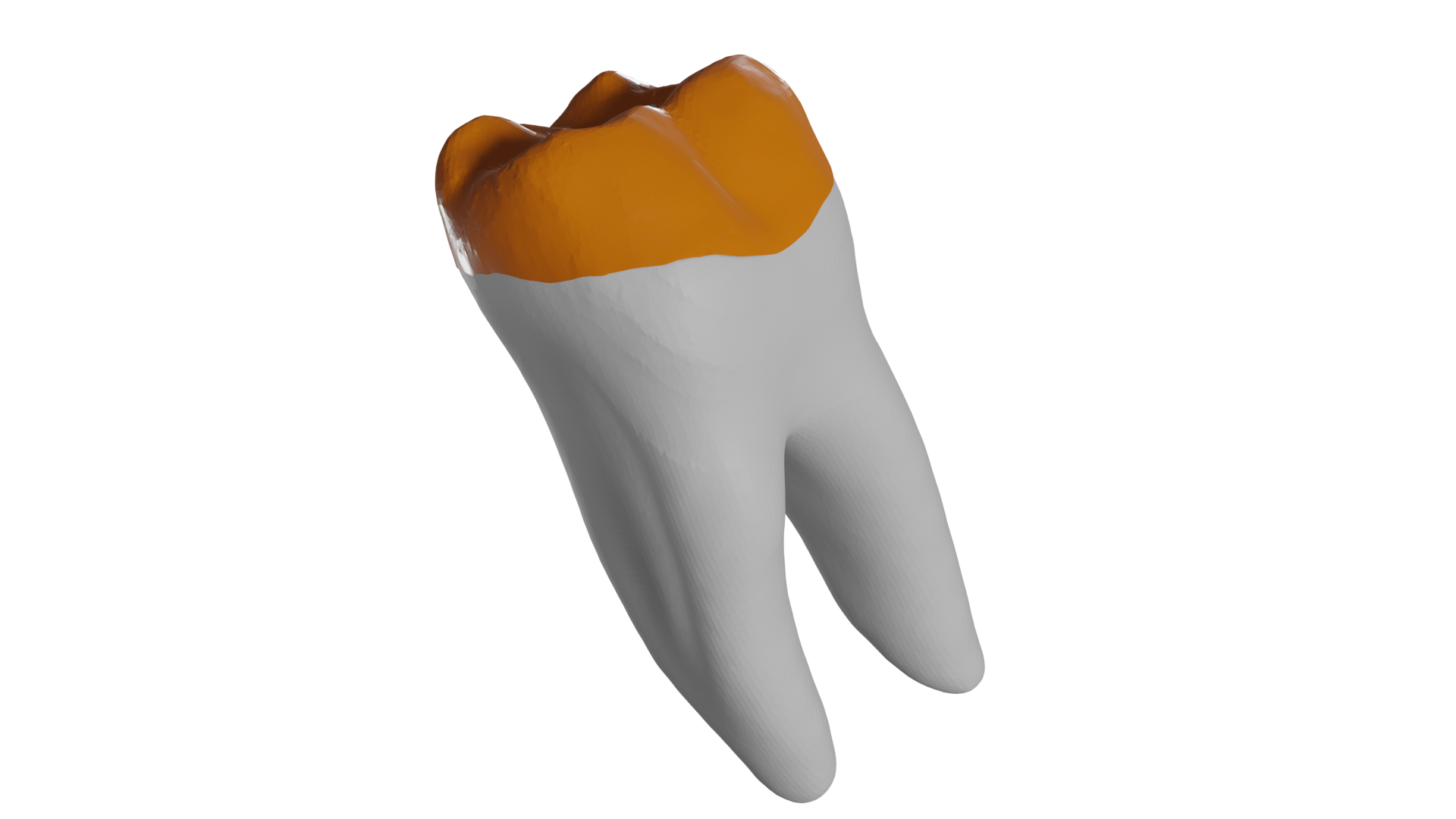
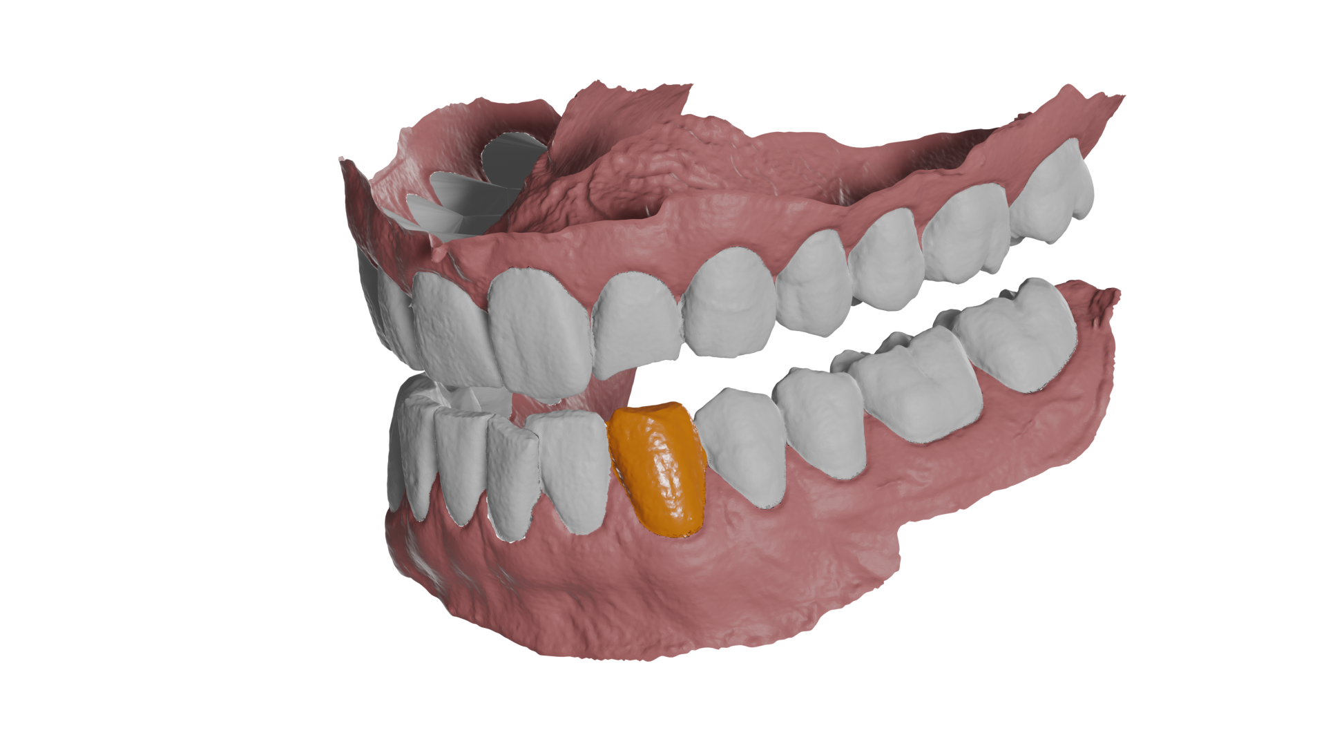
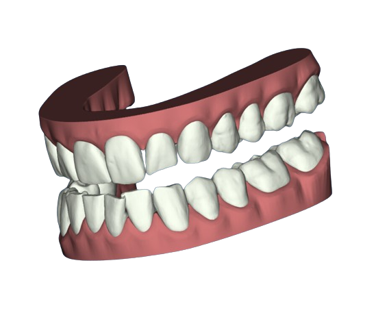
A single object that combines the crown of the Intra-oral scan with the roots from the Cone-Beam CT-scan. This results in the most accurate representation of the tooth possible.
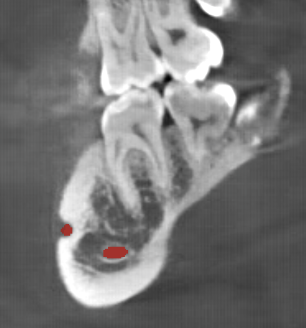
Automatic computation of the nerve canal paths in the mandibular bone.
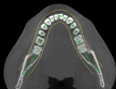
Automatic computation of panoramic arch on the CBCT scan.
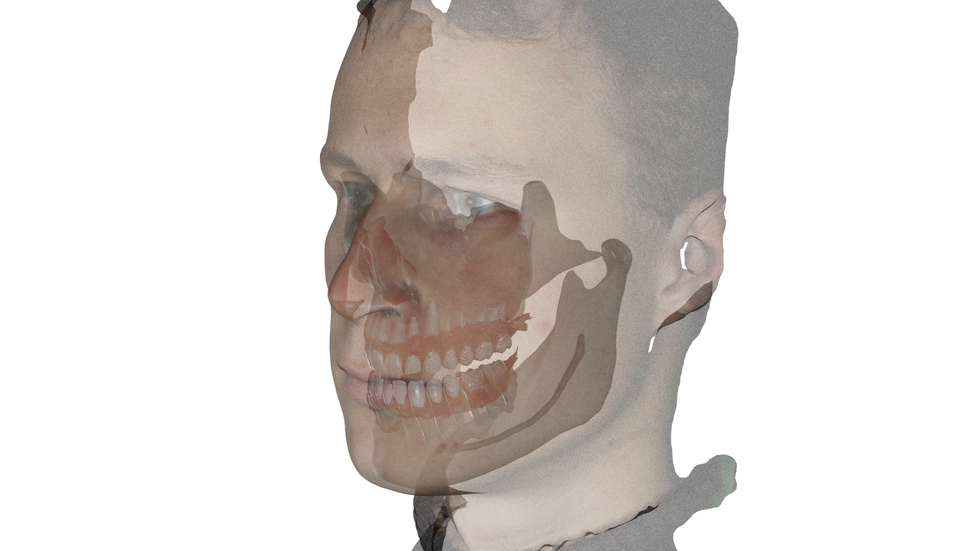
Automatic registration (matching) of the facial scan to the Cone-Beam CT-scan.
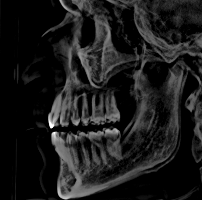
Automatic computation of the CEPH, Posteroanterior CEPH and panoramic projections based on the CBCT scan.
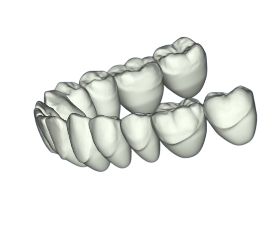
Automatic generation of closed crown meshes based on the intra-oral scan. The resulting meshes have a closed bottom, resulting in visually more appealing models.
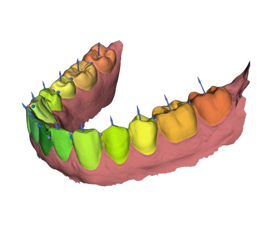
Automatic computation of key landmarks on the intra-oral scan. Currently returns the following landmarks for each segmented tooth:
- origin (center)
- tooth axis (long axis)
- mesial point
- distal point

Automatic base adding to the raw IOS.
Automatic generation of a closed gingiva base or emergent profile.
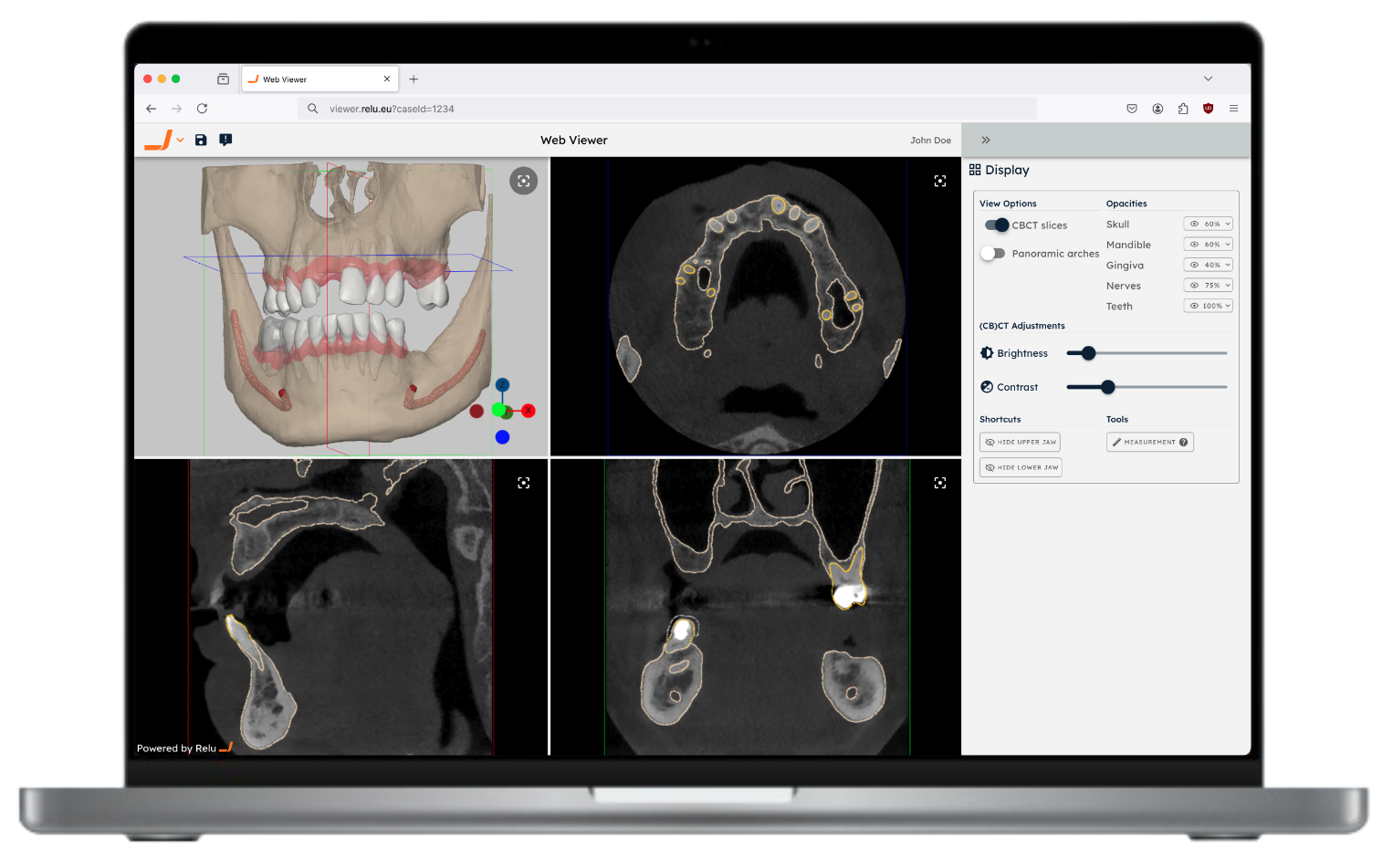
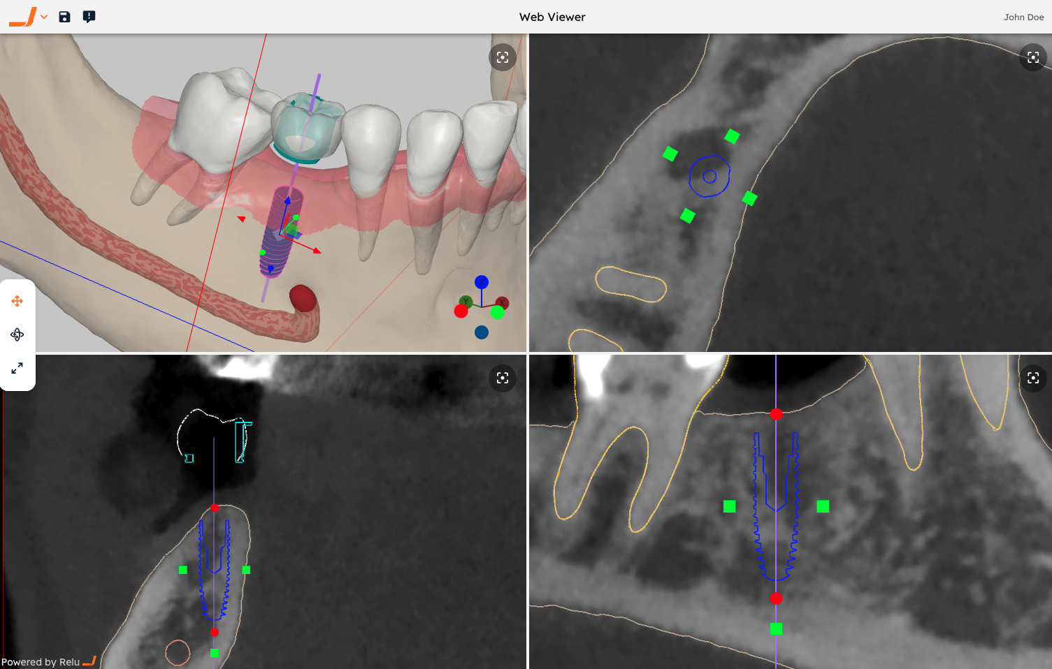
Ability to get back a URL to a web viewer, displaying the results in 2D and 3D, directly within the browser of your end-user. This viewing session is specific to your job (case) and can be easily shared with others, e.g. for quick inspection, for patient communication or for easy collaboration on a case.
The viewer is able to visualize most of the features listed above (e.g. the segmentations, mesh outputs, panoramic arches, ..). Additionally, some results will be able to be edited and exported (downloaded) directly from the browser window.
The UI of the viewer can optionally be branded to your needs, e.g. using your logo and color palette.
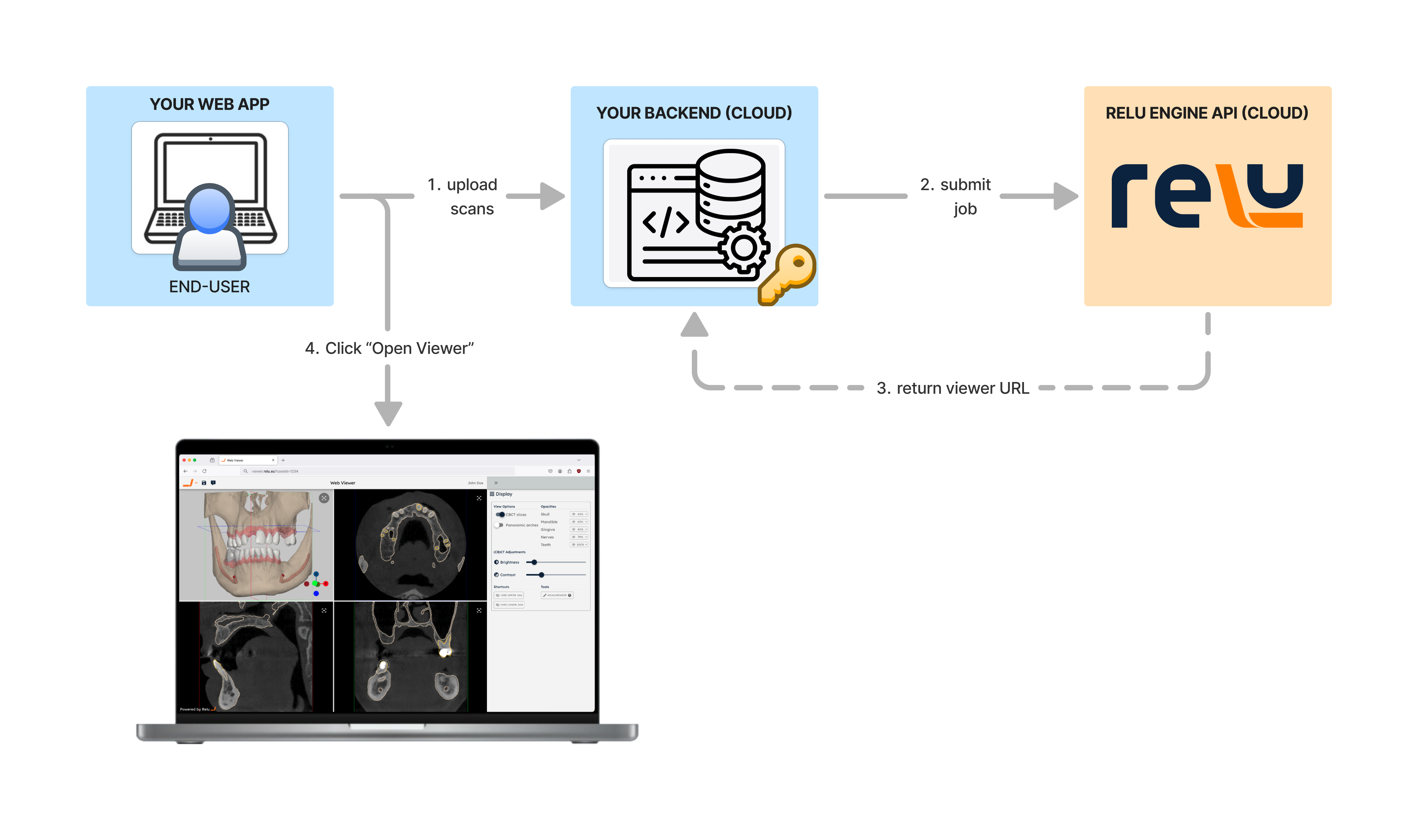
The diagram below illustrates a typical workflow, where the Web Viewer is integrated in your software. This way, your end-users are seamlessly redirected to the viewer, without the need to leave your application.
Finally, we are actively developing additional modules which can be added on top of the viewer:
- An implant module (shown on the right), allowing to visualize an automated implant planning proposal. Within the viewer, the end-user can inspect and alter the proposal, adjusting the position, orientation and dimensions of the implants, wax-ups, sleeves and surgical pins - both in 2D and in 3D. We are integrating several implant libraries.
- An orthodontic module, allowing to visualize an automated orthodontic setup proposal. Within the viewer, the end-user can inspect and alter the proposal, adjusting the position and orientation of the teeth.
Please contact us if you would be interested to integrate the Web Viewer in your workflow. We are still actively developing this feature and would love to hear your feedback.
More advanced options and outputs are possible. Find out more in the Designing your job body section, or get in touch with us for a custom solution.